
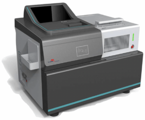
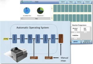
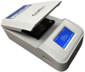
Efficiency and openingspan
• Total automation of the process (dewaxing, rehydration, permeabilization, enzymatic digestion, dehydration) with intermediate washes
• Extemporaneous enzyme preparation (automatic dilution from concentrated solution)
• Heating the slides at the start of the protocol
• Protocol standardization
• Suitable for all probes, regardless of supplier
Practicality
• Ready-to-use reagents (except enzyme)
• Ready-to-use enzyme dilution buffer for automatic extemporaneous preparation
• Management of 1 to 20 slides per run, with adaptation of the reagent volume used
• Management of multiple digestion times in a single run using semi-automatic mode
• Pre-recorded default programs
• Customizable programs
• French-language software interface with touch-screen control panel
• Monitoring of technique progress, with indication of time remaining in the current step and total time.
Operator safety and protection
• Reduce reagent handling by using ready-to-use solutions
• Use of substitutes for the dewaxing step
• “Autoboot” of the appliance on start-up with check of correct operation
• Self-cleaning of all manifolds on ignition and at the end of the process
• Control of reagent volumes drawn from and dispensed into the reaction chamber, with alarm in case of insufficient volume
• Waste volume control with fill alarm
• Presence control of waste, alcohol and distilled water containers with alarm
Dimensions and electrical features
• Floor-standing machine with adjustable feet and castors.
• Dimensions: 700 x 500 x 1000 mm (W x D x H)
• Weight: 50 kg
• Voltage: 220-240 V
• Power: 1500 W
Reagents not supplied
• Distilled water (800 to 1000 ml for a cycle of 20 slides)
• Absolute alcohol (300 to 500 ml per cycle of 20 slides)
Main functions
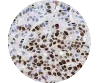
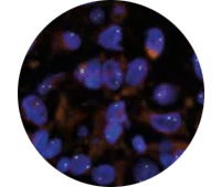
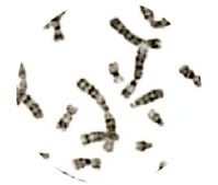
Makes in situ hybridization :
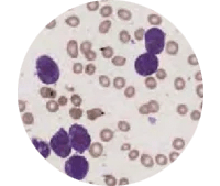
Staining process
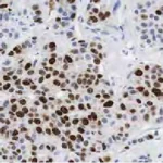
Areas of application
Features
Dimensions, weight, power
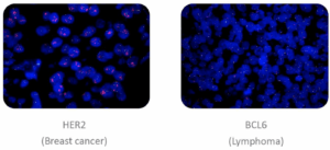
Since its creation, the company has advanced its fluorescently-labeled nucleotide production techniques from the design to the manufacture of FISH probes. To date, Cytotest has developed and successfully tested a highly diversified range of FISH DNA probes for molecular diagnostic applications in oncology, personalized medicine, prenatal testing, oncology and other fields.
The Cytotest range includes probes for both clinical and research targets, as well as customized products (custom probes). New products are constantly being added, partly thanks to our in-house R&D efforts, but more often as a result of requests from our customers to develop specific tests, particularly for rare cancers and companion diagnostics.
All products are manufactured in compliance with current regulations, and undergo quality testing to ensure they meet the most stringent standards. Regular optimization efforts have resulted in products of excellence. At the same time, Cytotest keeps a close eye on scientific and technological advances, observes cutting-edge discussions on diagnostic industry standards, certification and regulatory requirements, and remains attentive to changes in consensus concerning in vitro diagnostics and laboratory testing.
With a multi-disciplinary team of passionate and dedicated researchers, Cytotest can perform manufacturing at any scale and is able to offer solutions for a wide range of diagnostic test needs, from standard probe production to flexible custom design. Cytotest scientists have strategic collaborations to develop molecular diagnostic tests for new and rare pathologies, uncovered diagnostic niches and for commercialization in underserved regions of the world.
The mission of CytoTest Inc. is to transform human genomic discoveries into useful products, and to design, manufacture and validate genetic and clinical diagnostic reagents of the highest quality. Their main objective is to develop state-of-the-art, yet affordable, molecular cytogenetic solutions. Cytotest is dedicated to providing cost-effective products, proactive customer service and reliable technical support.
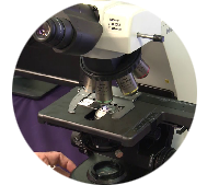
The machine offers a number of advantages, including convenient touch-screen programming, an integrated USB port, unrivalled heating speed and precision, minimal temperature variation and a higher humidity range. A unique design with liquid gutters provides convenient humidification and stable humidity control inside the chamber. The instrument is easy to clean, and there’s no need to use disposable foam strips for humidity, or any other consumables to replace.
Accepting a wide range of sample types, the tray is equipped with sliding guides that hold the slides in place and allow one-handed placement and removal. Its use considerably reduces working time without compromising precision and reproducibility.
The product is suitable for a wide variety of experimental denaturation and hybridization strategies, with four distinct modes of operation: denaturation/hybridization, hybridization, customization and PCR processes in situ. The integrated hot-water tank and sealed heating cover ensure perfect experimental reproducibility, for processing up to 12 slides simultaneously.
Available for 110-120V or 220-240V power supply.
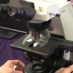
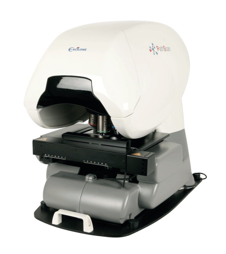
PathScan® Combi is a histological slide scanner that combines transmitted light and fluorescence scanning thanks to a dual-camera system (color and monochrome):
PathScan® Combi enables brightfield and fluorescence images to be matched for FISH analysis using EVA® software, in the same tumor zone as that previously identified by the pathologist in brightfield.
With its dedicated Web application, PathScan® Combi provides remote access to acquired images and password-protected access to individual files and working environments.
Specifications :
The scanner complies with the In Vitro Diagnostic Regulation (IVDR) (EU) 2017/746.
Allows you to centralize all your virtual blades in the same environment, regardless of image format.
Excilone® View Web can be installed on physical servers or in the cloud, and an SSL certificate with Https protocol ensures a secure connection and data protection.
The Excilone® View Web license is perpetual with no limit on the number of users.
Excilone® View Web is available in English and French. The platform is accessible from PCs, Macs and tablets. And it’s browser-compatible.
Excilone® View Web allows you to perform the following operations:
Key words :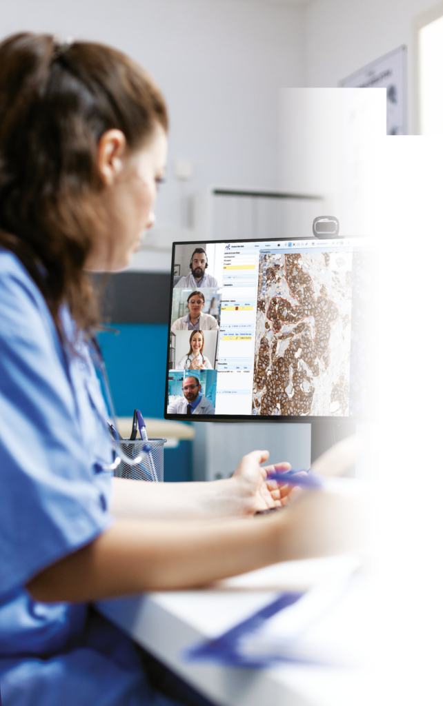
Compatible image formats :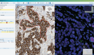
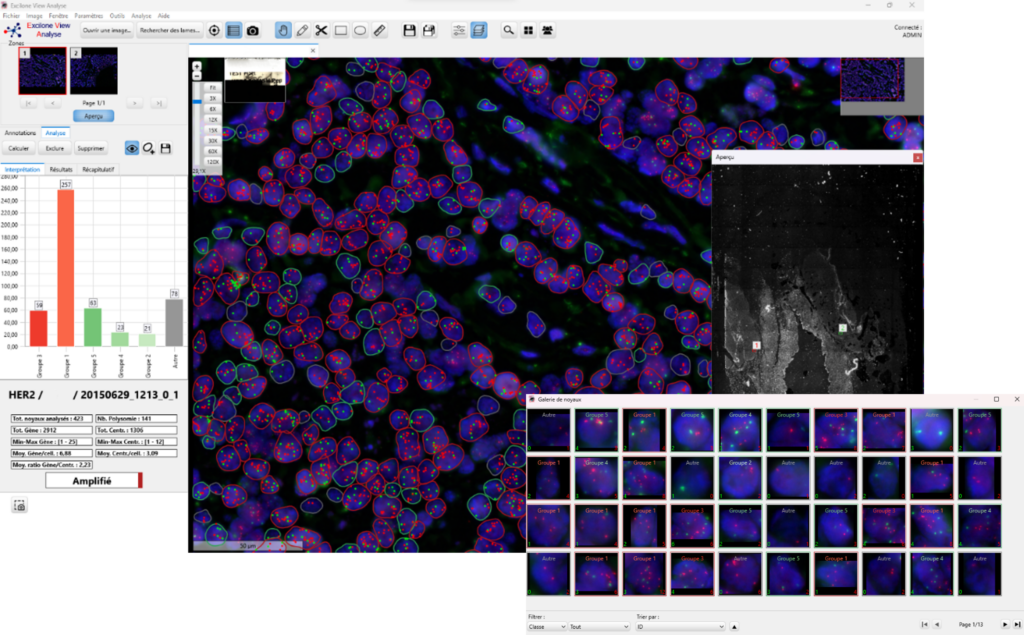
EVA® offers advanced features for intuitive navigation of all your slides (brightfield, fluorescence).
With Excilone® View Analyse you can also :
The benefits:
For research only