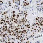

The construction of Tissue Micro-Arrays (TMA) is a tedious task with many steps that are prone to errors.
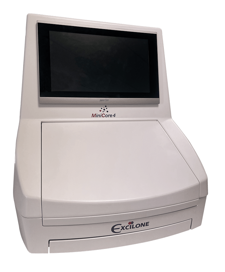
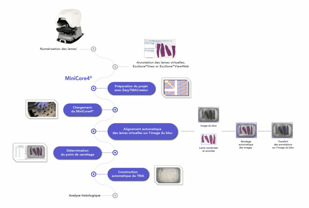
MiniCore® 4 greatly simplifies operations by automating the construction of Tissue Micro-Arrays. The MiniCore®4 software displays all the real-time information. It performs the superposition and the registration automatic creation of virtual slides annotated on the image of the donor blocks using artificial intelligence, which allows very precise selection of coring areas.
MiniCore® 4 features a large loading capacity with 4 blocks receivers and 16 donor blocks per board. It also offers 4 games needles ranging from 0.6 mm to 2.0 mm in diameter.
Compact and ergonomic system
Advanced technology
Optimal traceability
Extended compatibility
Excilone®View software allows you to view and select areas of interest on scanned slides. Excilone®View is compatible with most slide scanners currently on the market. Try the free version of Excilone®View (Digital Pathology – Excilone).
Precision of tissue sampling
Large capacity
|
Description |
Reference |
|
0.6 mm “donor” needle |
MIN04-06D |
|
0.6 mm “receiver” needle |
MIN04-06R |
|
1.0 mm “donor” needle |
MIN04-10D |
|
1.0 mm “receiver” needle |
MIN04-10R |
|
1.5 mm “donor” needle |
MIN04-15D |
|
1.5 mm “receiver” needle |
MIN04-15R |
|
2.0 mm “donor” needle |
MIN04-20D |
|
2.0 mm “receiver” needle |
MIN04-20R |
* Optional steps: only when the process is performed with scanned slides.
CE Marking : Built to CE/EMC requirements and certified to these standards.
|
Component |
Description |
|
Dimensions |
Width : 560 mm Depth : 720 mm Height : 650 mm |
|
Weight |
Approximately 45 kg |
|
Alimentation |
85/264 V~, 47-63 Hz, 150 VA |
|
Number of receiving blocks |
up to 4 |
|
Number of donor blocks |
up to 16 |
|
Available needle sizes |
0.6 mm / 1.0 mm / 1.5 mm / 2.0 mm |
|
Work surface on the block |
40 mm x 28 mm |
|
Thickness max. thickness |
10 mm |
|
Length of carrots |
Configurable on the software |
|
Coring area |
Manual determination on the block image. |
Indicative but non-contractual specifications.

MiniCore®3 includes an 8-position carousel as standard, allowing you to use up to 7 donor blocks simultaneously, saving you even more time and effort.
Core zone selection in donor blocks is performed by zone drawing or pointing, directly on screen on the donor block image superimposed on that of the H&E control blade.
The MiniCore®3 Control Station software interface displays all information in real time. In the event of difficulties on a donor block, the user can easily switch blocks and continue his tissue array, without any loss of quality.
With our TMA construction software, you can design any type of format from a list of Excel® specimen identifiers.
During construction, MiniCore® 3 automatically uses your tissue array configuration file. Position errors are virtually eliminated.
Whenever a donor block is required, its ID is displayed on the screen. Specimen errors are greatly reduced.
Finally, each step is recorded in a log file to guarantee perfect traceability.

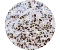
L’identification des spots est certainement l’étape la plus consommatrice en temps opérateur et l’étape la plus difficile pour la préparation des spots de tissue Arrays en vue de leur analyse. L’identification inclut la détection des spots sur le scan de la lame puis l’association de chacun de ces spots avec leur position respective dans le plan initial de votre tissue Arrays afin de créer le lien de traçabilité essentiel entre le spot et les données patient.
Spot Browser® 4 inclut un outil de de-Arraying qui permet la détection et l’association des données. Il vous suffit d’importer votre fichier de la carte de votre tissue Array et Spot Browser® 4 s’occupera du reste sans efforts. Des interactions et corrections sont toujours possibles dans les cas où le tissue Array est véritablement en mauvaise condition sur la lame (fortes déformations).
Dans de nombreux cas, les spots sont “scores” visuellement parce que l’oeil expert du pathologiste ne peut pas aussi facilement être remplacé par un logiciel aussi puissant soit-il.
Pour répondre à ce besoin, Spot Browser® 4 permet à l’utilisateur de créer ses propres champs de données sous divers formats (libre, numérique, liste déroulante…) pour saisir ses annotations. L’utilisateur peut ainsi très rapidement naviguer d’un spot à un autre et saisir les annotations.
Les données saisies et les données importées peuvent être exportées sous format Excel pour traitement statistique.
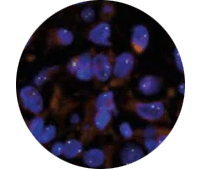
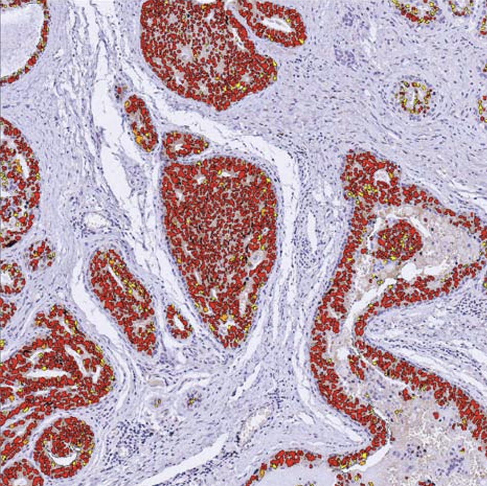
Pour l’analyse d’un grand nombre de spots ou pour des analyses quantitatives, Spot Browser® 4 propose un outil très complet pour l’analyse d’image automatique.
Les caractéristiques exploitées sont couleur HSV et morphométrie. Chacune de ces caractéristiques peut être utilisées seule ou en combinaison suivant les objets à détecter.
L’utilisateur peut très aisément créer ses propres protocoles de détection avec un ou plusieurs types d’objets à détecter et avec ou non imbrication pour permettre notamment de comptés des noyaux marqués dans une zone tumorale. La détection de marquage cytoplasmique et membranaire est également possible.
Les protocoles de détection et d’analyse peuvent être sauvegardés dans des librairies internes pour pouvoir être réutilisés ultérieurement.
L’utilisateur peut analyser les spots un à un ou en batch automatique et les analyses peuvent combiner des interventions humaines (traçage de zone par exemple) avec des modes complètement automatiques.
Les résultats en données brutes et interprétées peuvent être exportés vers Excel.
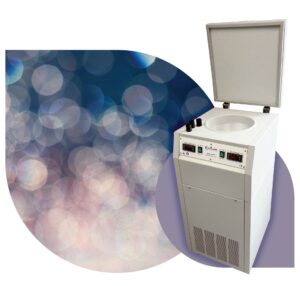 Le SnapFrost® garanti une congélation ultra rapide et reproductible
La technique de référence utilisant l’Iso pentane réfrigéré jusqu’à -80°C, non dégazant contrairement à l’azote liquide, permet une transmission rapide de la température à l’échantillon de façon reproductible.
Le contenu moléculaire ne diffuse pas dans l’échantillon, les membranes cellulaires sont conservées et les lésions tissulaires sont évitées.
Ergonomique et souple d’utilisation, le SnapFrost® dispose de deux chambres de congélation, contrôlables individuellement, pour s’adapter à tous les protocoles de congélation et les types tissulaires. Le volume de la chambre basse contenant l’Iso pentane peut être modulé (de 5 à 680 ml) pour s’adapter à toutes les tailles d’échantillons.
Les solutions alternatives comme le Novec 7100 peuvent également être utilisée dans le SnapFrost®, en conservant les avantages de l’appareil et en garantissant une haute qualité de congélation.
Programmable, compacte et autonome, le SnapFrost® peut être déplacé sur les différentes zones de congélation, vous garantissant flexibilité, gain de temps, sécurité, qualité et reproductibilité dans vos opérations de congélation.
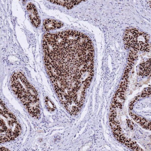
Les contrôles tissulaires traditionnels ont longtemps été la norme en immunohistochimie mais ils présentent des limites : manque de standardisation, variabilité d’un lot à l’autre et risque d’utilisation abusive. Les lames PRS révolutionnent l’immunohistochimie en remplaçant ces contrôles tissulaires.
La standardisation du processus de coloration IHC à l’aide de l’outil PRS offre plusieurs avantages dans la validation des anticorps :
Une évaluation quantitative : la quantification prédéterminée des antigènes de substitution permet une évaluation quantitative de l’intensité de la coloration, facilitant ainsi l’évaluation objective de la performance des anticorps utilisés.
Un contrôle qualité : l’outil PRS sert de mesure de contrôle de la qualité, permettant aux utilisateurs de vérifier la fiabilité et la cohérence de la coloration des anticorps entre différents lots et expériences.
Une détection des erreurs : les écarts entre le modèle de coloration observé et le résultat attendu basé sur l’outil PRS indiquent des erreurs potentielles dans la sélection ou l’application des anticorps, ce qui incite à des recherches plus approfondies et à des mesures correctives.
Efficacité du flux de travail : la standardisation du processus de coloration rationalise les procédures de flux de travail, réduit la variabilité et optimise l’efficacité du laboratoire dans les tests IHC ; plus de motif de tissu contrôle à déposer sur la lame en plus du tissu à tester.
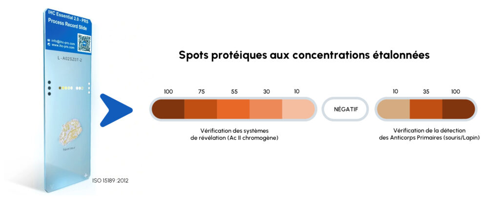
PRS élimine les résultats de coloration faussement négatifs/positifs grâce à un contrôle à 100 % sur lame ; chaque lame IHC est évaluée individuellement.
PRS permet de s’affranchir des blocs contrôles du commerce, onéreux.
PRS garantit des résultats précis et cohérents tout en respectant les normes ISO 15189. Les cibles protéiques primaires et secondaires avec des niveaux d’expression antigénique certifiés sont reconnues comme un dispositif médical ISO 13485. PRS aide à obtenir l’accréditation et la conformité réglementaire aux normes ISO 15189, CLIA et ACP.